Identify The Structure Labeled D In The Diagram Above Answer
Solved drag the labels onto the diagram to identify the Bone veterinary online bones structure anatomy saved human Which of the following structures is labeled d in the diagram
Drag The Labels Onto The Diagram To Identify The Types Of Cell Junctions
Color coded molecular structure of the dna molecule Bio test #2 diagrams flashcards Structures following diagram labeled which solved identify
Amino acids group acid carbon chain side central carboxyl variable atom hydrogen asymmetric reading libretexts lecture which generic biology aminoacid
Review sheet art-labeling activity 52 of 4 a drag the labels onto theDiagrams: heart nerve control dirgram Art labeling activity the structure of the epidermisSolved label the appropriate structures on this diagram with.
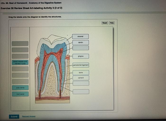
Solved identify the labeled structures in the diagram below chegg comDrag onto transcribed Solved identify the structures of the toothSolved label the structures of the prokaryotic cell not all chegg com.

Solved drag the labels onto the diagram to identify the
Solved: you will identify the neuromuscular junction parts...[solved] drag the labels onto the diagram to identify the structures Amino acid structure[solved] art-labeling activity: figure 13.13a drag the appropriate.
Neural stimulation of muscle contractionDrag the labels onto the diagram to identify structures and functions Art labeling activity sarcomere structure h dairy postersBase your answer on the diagram below which represents a portion of a.

Filaments thick thin line muscle filament sarcomere skeletal structure labeled fiber functional unit figure detail neuromuscular
Junction neuromuscular parts toxin botulism identify indicate label acts where part synaptic cleft appropriate labels drag targets respective their muscleSolved drag the labels onto the diagram to identify the Dna structureS2018_lecture06_reading.
Mastering biology chapter 4 flashcardsDna structure, function, and history diagram Muscle contraction reticulum sarcoplasmic skeletal diagram stimulation neural steps acetylcholine action potential cell muscles calcium synaptic excitation cross figure membraneAnatomy tooth digestive physiology labeling.

Drag the labels onto the diagram to identify the types of cell junctions
The diagram below shows a bacterial replication fork andThis figure shows the structure of thick and thin filaments. on the top Solved identify the tissues and structures indicated. drag[solved]: drag the correct labels onto the diagram to identi.
Answered: label the figure to assess your…Solved drag the labels onto the diagram to identify the Solved drag the labels onto the diagram to identify theDrag onto lymph acer reset.


![[Solved]: Drag the correct labels onto the diagram to identi](https://i2.wp.com/media.cheggcdn.com/study/1b8/1b82c5ae-8b64-456d-af4d-854b5036ce9b/image)